Minimally Invasive Breast Biopsy
Breast cancer is now the most common cancer among women in many countries. The incidence of breast cancer is rising and this has been attributed to a combination of multiple factors including modernization of society, changing lifestyles, women bearing children later and shorter duration of breastfeeding. Leading a healthy lifestyle may reduce a woman’s risk of breast cancer. However there is no absolute prevention and the best strategy to beat breast cancer is early detection and treatment. Breast cancer is one of the most treatable cancers with excellent survival rates when treated early, so it is crucial to clinch the diagnosis in the early stages.
Breast Cancer Screening
Screening is the detection of abnormalities or disease before the onset of symptoms. Breast cancer screening is important as it allows the cancer to be detected in its early stages.
Women should practise breast self examination once a month to look out for lumps, abnormal discharge or skin changes. Annual mammogram screening is performed for women 40 years old and older. For younger women or in women >40 years old with dense breasts, the option of ultrasound screening should be discussed. Ultrasound is helpful in detecting abnormalities that may be missed if the breasts are dense, and can also be used to provide more information if an abnormality is detected on mammogram.
Minimally Invasive Breast Biopsies
When a lump or abnormality is detected, the most definitive way to determine the nature of the lump is to perform a biopsy. A biopsy means removal a sample of the lump, or even the whole lump for analysis. There are different types of biopsies available including fine needle aspiration, core needle biopsy, Mammotome® (vacuum assisted) biopsy, radiofrequency INTACT® biopsy and open surgical biopsy. The last three methods of biopsies remove the entire lump or abnormality, with the Mammotome® and INTACT® biopsies being minimally invasive.
A) Fine Needle Aspiration Biopsy
This is a very simple form of biopsy that can be done in the clinic and only takes a few minutes to perform. A fine-bore needle (similar to that used for blood tests) is inserted into the breast lump and 1-2 drops of material is aspirated from the lump. The material is spread on a slide and the cells will be analysed by the pathologist. This method is also used for draining cysts that are large or symptomatic. It is important to understand that while this method is >90% accurate, occasionally there may be false positive and false negatives, so a benign result will still require close follow up of the lump.
B) Core Needle Biopsy
Core needle biopsy studies architecture of the lump by extracting small fragments of the lump for analysis via a small puncture wound in the skin. Hence compared to the fine needle aspiration biopsy, it is much more accurate. It is done under local anaesthesia and takes about 15-30 minutes to perform, either in the clinic or hospital setting. It is usually done under image guidance eg ultrasound guidance. This is the most common type of biopsy performed for highly suspicious breast lumps.
C) Image Guided Mammotome® Biopsy
Mammotome® biopsy is also known as vacuum assisted biopsy. In this method, a hollow needle is inserted into the breast under ultrasound or mammogram guidance to the area of the breast abnormality. When activated, the needle with the aid of vacuum suction and blade is able to remove the entire abnormality in multiple cores. Breast lumps of up to 2-2.5cm can be removed completely using this method. The mammotome® biopsy is a versatile method that can be used in the majority of patients. This is one of the most common biopsies performed for abnormal mammogram microcalcifications and small indeterminate lumps detected on ultrasound. It is usually performed under local anaesthesia.
D) Minimally Invasive Image Guided Radiofrequency INTACT® Biopsy
This is a relatively new form of biopsy technique that uses a radiofrequency enabled wand or needle probe. The wand is inserted through a small incision in the skin and directed towards the breast lump under ultrasound or mammogram guidance. A “basket” is then activated and captures the whole lump using radiofrequency. The lump is then removed through the small skin incision as a single tissue specimen. In selected cases, removal of the lump as a single specimen may be preferable with advantages for pathological analysis. Lumps of up to 2.0cm may be successfully removed with INTACT® and there is less risk of bleeding in this method. However, it may not be suitable for all patients depending on the location of the lump and distance of the lump from the chest wall and skin.
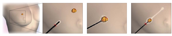
INTACT® wand with capture and removal of the breast lump
Both these methods of minimally invasive breast biopsies can be safely and comfortably performed under local anaesthesia and take about 15-30 minutes. An unsightly scar from open surgery can be avoided as these methods only result in small scars 3-6mm in size depending on the size of the lump and size of the needle or wand used.
Conclusion
Breast cancer is highly treatable with excellent rates when detected and treated early. There are many forms of minimally invasive breast biopsies now that enable a definitive diagnosis without surgery and unsightly scars. A definitive diagnosis allows your doctor to plan your treatment, such as further surgery or chemotherapy for cancer. If you have a mammogram or ultrasound abnormality, do consult your doctor regarding your suitability for a minimally invasive breast biopsy. Remember that a biopsy provides confirmation of the diagnosis, peace of mind and in some cases early institution of treatment as and when necessary.
Prepared by:
Dr. Tan Yah Yuen
Breast Surgery
BreastCare Surgery Pte Ltd
Disclaimer: The views and opinions in the article are the writer’s own and do not necessarily reflect those of Mount Elizabeth Medical Centre (MEMC). The writer is fully responsible for the accuracy, completeness and usefulness of the information provided in the article. MEMC will not be liable for any errors, omissions or copyright issues with regard to the contents of the article.